
How 3-D Printing Is Revolutionizing Medicine
By: Hammad Khalid
3-D printing is the process of making a three-dimensional solid object from a digital model. This futuristic process first began in the 1980s. Recently, the 3-D printing market has grown into a multibillion-dollar industry due to substantial price drops and widespread commercial availability of 3-D printers. This resurgence in popularity has led to multiple novel applications for 3-D printing, including jewelry, art, footwear, dentistry, civil engineering, architecture and education. Perhaps the most creative use of 3-D printing lies in biomedical engineering and its applications in healthcare such as bioprinting.
Bioprinting is a relatively new process that uses 3-D printing technology to recreate functioning, viable body tissue by dispensing cells on a biocompatible material using a layer-by-layer approach. Using this technique, biomedical engineers have begun printing prototypes of many different body parts including heart valves, ears, joints, skin grafts and artificial bone.
How did this phenomenon begin?

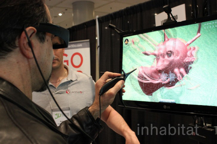
Photo credit: Inhabitat / Foter / CC BY-NC-ND
In 2000, biomedical engineer Thomas Boland filled his Lexmark printer’s ink cartridge with collagen and fed the printer a black silicon sheet onto blank paper. After successfully printing his initials with off-white proteins, he moved on to printing with E. coli bacteria, then larger mammalian cells from lab rats and Chinese hamsters. 90 percent of the cells remained viable after printing, which made for an incredibly useful product. Boland filed his first patent for printing cells in 2003.
While Boland laid the foundations for 2-D bioprinting, other biomedical engineers tackled synthetic medical challenges in 3-D by printing dental crowns from porcelain, hearing aids from acrylic and bone grafts from ceramic. As these same techniques paved the way for 3-D bioprinting, its applications began to increase exponentially in both form and function. Soon, scientists began experimenting with different types of “ink” for their bioprinters, as is the case with Cornell engineer Hod Lipson. Lipson was the first to utilize bioprinting to create cartilage, which he then used to print a meniscus to replace the cushioning in the knee.
Bioprinting has already begun saving lives. Scott Hollister, a biomedical engineer who runs the University of Michigan’s 3-D Printing Lab, has created a type of splint to hold open the windpipe until it is strong enough to work on its own. Hollister, working with Dr. Glenn Green at the University of Michigan, has already used this device successfully on two babies with tracheomalacia. In addition, Hollister has built a jawbone for a patient in Italy.
However, there are still many roadblocks biomedical engineers face in the process of converting their synthetic lab creations into real, feasible body parts ready to transplant into human bodies. In Lipson’s case, his meniscus was deemed to be too weak to endure routine wear-and-tear by knee-replacement surgeons. Different body cells have different functions and therefore must be made with certain specific requirements depending on the type of tissue they become. To create an artificial meniscus, for example, one might need to develop a bioreactor that can use heat, light or auditory pulses to mold the tissue into proper formation.
While bioprinting is still a new, developing technology, companies like Organovo have already made substantial progress in the field. Scientists at Organovo have built cardiac tissue by stacking piles of cells along the z-axis, which, when fused, have beat in unison like a normal heart. Bioprinters are being used to form tubular structures by depositing layers of hydrogel rods and bio-ink consisting of thousands of human cells. After printing, the hydrogel rods are removed from the tubular structures to form blood vessels. These vascular grafts can be combined with cardiac, liver or lung tissue to form complex organs. Organovo’s first marketable product is liver tissue for clinical trials. Currently, liver toxicity is the number one reason for a drug to be pulled from clinical trials, as well as off the shelves after post-market approval, according to the Food and Drug Administration. Even animal trials, which are the predominant method of testing for liver toxicity today, are not a reliable approach to test how drugs will affect the human liver once ingested. Organovo’s liver assays could expedite the presently prolonged clinical trials drug testing process.
So what are some promising future applications for such a groundbreaking technology like bioprinting? The ultimate challenge of producing a whole, fully-functioning, transplantable organ still remains. The national organ donor waiting list currently rests at over 118,000 people and grows by over 300 people each month. Naturally, the odds of finding a donor match are low. Bioprinting organs with cells made from a patient’s own body would be a perfect solution. Instead of training with cadavers, surgery residents could practice on bioprinted organs with prebuilt tumors or defects before operating on cancer patients.
Researchers at Princeton University have been experimenting with bionic organs that not only repair but also extend human abilities. For instance, they created a matrix of hydrogel and bovine cells in the shape of an ear and incorporated silver nanoparticles, creating a silver antenna that could pick up radio frequencies beyond the range of normal human hearing.
These recent discoveries in the field clearly show that bioprinting is at the forefront of medical innovation and is here to stay.